
Dr. Leon Wortham
Dr. Leon Wortham is the Owner, President & CEO of Wortham Laboratories, Inc. (Tennessee), which Develops new products (diagnostic, medical devices, pharmaceuticals), improves existing products (SeraSeal™ has CE Certification). He has previously worked as consultant for biotech companies. As R & D Chemist at Medical Analysis Systems, Camarillo, CA, Dr. Leon developed new clinical products, wrote SOPs and Quality Assurance procedures, and Hexcel Corporation, Chatsworth, CA where he Developed polyurethanes for medical applications, NASA project, S Military missile system. At Bio-Vet Laboratories, Annapolis, MD, as CEO he formed a veterinarian reference laboratory, developed clinical controls and reagents, supervised a team of 32 employees.
From 1978 – 1998, he worked in Trauma/Emergency Hospital Centers – Level 1
Dr. Leon Wortham is a graduate from CLA School of Medicine, Westwood, CA, in Trauma Surgery His Research History includes Investigational Studies: Multiple Sclerosis, from UCLA School of Medicine, Reed Neurological Research Center and credits with 43 years industrial research and development in Products: diagnostic, medical devices, pharmaceutical & Specialty (isolating, purifying, stabilizing proteins, enzymes, hormones). He has 7 patents to his credit. His publications include contributions to J. Neurochemistry, 1975, “Acid Proteinase: Pinpointing the Source, Using Experimental Allergic Encephalomyelitis as a Model.” and American Association of Clinical Chemists; American Society of Hematology
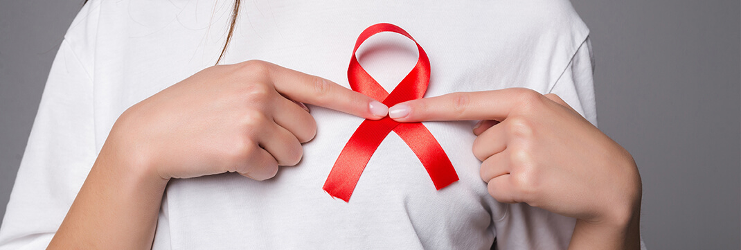
The birth of SeraSeal™, the world’s first and only primary Hemostatic agent!
How did Dr. Leon Wortham conceptualize SeraSeal™?
“One evening, while working at a Level 1 Trauma Hospital in Los Angeles, the paramedics brought in a gunshot case to the chest. The patient was bleeding profusely, and we had no choice but to open his chest in the trauma unit. He had sustained a wound from a 38-caliber bullet to the arch of his aorta. We did everything we could to control the bleeding, but the patient was already in a state of coagulopathy due to shock. As a result, the patient died at the age of 31, leaving a wife and two small children. Up to that time, I had seen thousands and thousands of patients who died from hemorrhage, and I was incredibly angry that we did not have a tool to arrest a Level 4 type bleed.
Early the next morning in my private laboratory, with my clinical and chemistry background, I formulated a single component, with the active agents and stabilizers in their appropriate amounts for a primary hemostatic agent, and it achieved hemostasis within in 1-2 seconds for normal whole blood, 3-5 seconds for anticoagulant (Heparin, Coumadin, aspirin) whole blood, with no change in clotting time after repetitive freeze-thaw cycles.”
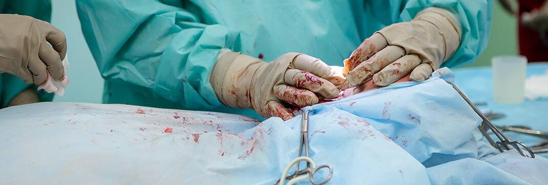
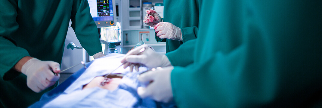
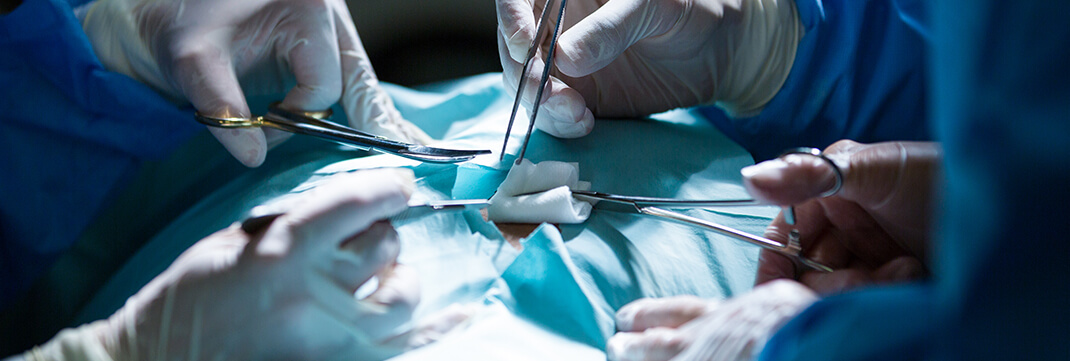
Surgical Procedures Accomplished
Lorem Ipsum has been the industry’s standard dummy
Brain Stereotactic Canal
The Todd-Wells stereotaxic guide was used in this procedure. The scalp entry point was infiltrated with 1% lidocaine with 1:100,000 epinephrine and a 1 cm incision were made. SeraSeal™ was applied and achieved hemostasis within 1-2 seconds. The arc system was attached to the base ring on the patient. A long 4.5 mm twist drill bit was used to make the craniotomy on the left side of the patient, at the coronal suture in the sagittal plane of the ipsilateral pupil. The twist drill guide tube in the arc slide bushing was exchanged for a 2.7 mm guide tube. A B-gauge cannula was then advanced to a depth of 7 cm at the skin-edge, toward the medial canthus of the ipsilateral eye and anterior to the targus. A blunt stylet was removed, and the biopsy forceps was advanced to the predetermined set point and biopsies obtained.Read More

Gastroenterology Subtotal Gastrectomy
A chevron incision was made to the abdomen, using Bookwalter retractors. Dissecting began by dividing the omentum from the transverse colon, using electrocautery. The lesser sue was entered, allowing assessment of the retroperitoneum, with regard to the tumor extension and lymph node involvement. The distal porti8on of the gastrectomy was performed. The origin of the right gastric artery at the common hepatic artery was identified, dissected, and SeraSeal™ was applied to the arterial bleed, achieving hemostasis in 5-8 seconds. After hemostasis had occurred the vessel was ligated with 2-0 silk sutures. Next the right gastroepiploic artery was identified and traced back to the gastroduodenal artery, which was similarly dissected, treated with SeraSeal™ with the same time to hemostasis, before being ligated. Read More

Craniotomy-Cranioplasty
A scalp incision was made directly in front of the targus curved posteriorly above the helix of the ear, and continued in a question mark fashion from the parietal area towards the frontal midline, ending behind the hairline. SeraSeal™ was applied to the scalp bleeding capillaries, achieving hemostasis in 1-2 seconds. Osteotomy was performed to reverse the right temporal bone. Multiple slit openings in the dura over the area of maximal clot thickness was performed, followed by irrigation of the hematoma and cerebral contusions with suction and cup forceps, facilitated by copious irrigation. The subdural space was explored. No resection to the temporal pole was necessary. SeraSeal™ was applied to several cerebral bleeds, arresting the bleeds in 1-2 seconds. In addition, SeraSeal™ was applied completely over the contusion area to avoid recurrent hematoma formations. Read More
Left Hepatectomy
The patient was placed in a supine position with the right arm extended at a right angle to the body. A bilateral subcostal incision extended in a vertical manner to the xiphisternum was made. The hepatic pedicle outside the liver was encircled with a vessel loop to facilitate occlusion during the liver resection. The gallbladder was dissected away from the liver. The lowermost hepatic veins behind the liver were dissected. SeraSeal™ was applied in this are due to a considerate amount of hemorrhage produced during the caudate hepatotomy. Hemostasis occurred in 2-5 seconds. Incisions were made in the liver capsule in two areas. The first was made in the caudate process immediately parallel to and to the right of the inferior vena cava. The full thickness of the caudate process was divided using diathermy crushing and ligation. Read More
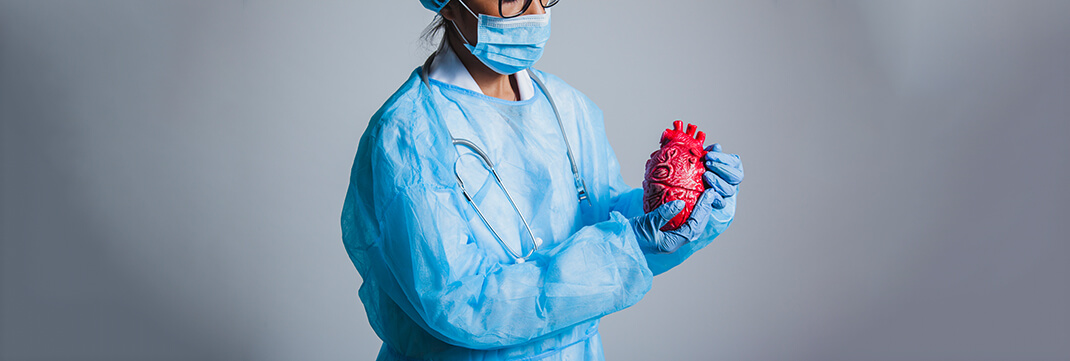
Heart Transplant
A median sternotomy was used to perform a thymectomy and to expose the patient’s heat. SeraSeal™ was applied to the marrow of the sternum, as well as the innominate and thyroid veins, achieving hemostasis in 1-2 seconds. The internal and inferior and superior arteries were ligated. Single venous and arterial cannulation was used. A standard orthotopic technique using biatrial connection was used. Prior to performing the transplantation, a tricuspid annuloplasty on the donor graft was performed. All aortic arch vessels were isolated with loose tourniquets during the initial cooling phase in preparation for reconstruction. Implantation of the autograft was accomplished with systemic hypothermia, performing the atrial anastomosis under low-flow perfusion, with the pulmonary artery clamped and systemic perfusion maintained by means of the arterial cannula positioned in the ductus arteriosus. Read More
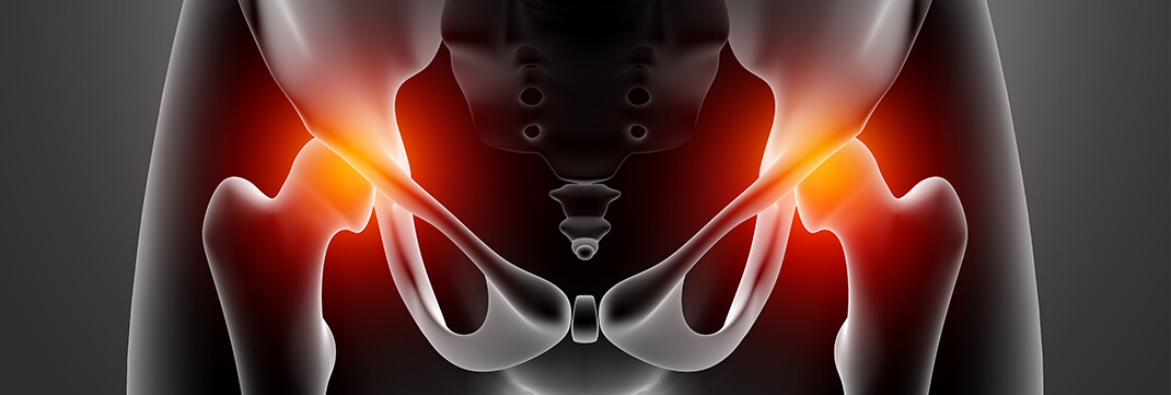
Primary Total Hip Replacement
Prior to surgery a template was used to measure sizing for the prosthesis. The stem size was 13 mm and the cup size to be 52 mm. The patient was placed in a lateral position, and an incision was made from the posterior superior iliac spine, then to the greater trochanter, and continued distally along the femur. SeraSeal™ was applied along the length of the incision, achieving hemostasis in 1-3 seconds. The fascia lata and the gluteus maximus fascia were divided in lieu of the incision. The maximus was split along its fibers. The gluteus maximus on the femur was completely divided to increase exposure. The short external rotators were incised close to the greater trochanter. The gluteus medius and minimus were subperiosteally raised from the ilium and retracted with sutures. The femoral head was dislocated by flexing and rotating the femur. Read More

Tonsillectomy Intracapsular Coblation
The patient was placed in the Rose position with a shoulder roll. A mouth prop was inserted, opened, and suspended. An Alysa clamp to the tonsil was used to allow for traction during dissection. Curettes were utilized to dissect the tonsils, and SeraSeal™ was applied to arrest the bleeding within 1-2 seconds.

Hysterectomy
A transverse incision was made 2 cm above the symphysis pubis, and the incision was taken down to the rectus fascia. The rectus fascia was dissected bluntly away from the underlying rectus muscle. The rectus muscle was separated in the midline, and the peritoneum was opened in the vertical midline. Using a Kocher clamp, the uterus was pulled up into the wound. The round ligament was suture ligated and cut. An avascular area in the broad ligament was bluntly fractured with a finger, producing an opening below the ovarian ligament and fallopian tube. Both the fallopian tube and ovarian ligament were clamped, cut, and ligated. Upward traction was placed on the uterus. The peritoneum in the anterior culde-sac was opened between the ligated round ligaments. The bladder was mobilized by dissecting it free of the anterior surface of the uterus and cervix. Read More
Mastectomy
Borders of the transverse incisions across the chest wall were established and an 8 mm thick flap extending 1 cm from the skin edge was developed. The plan between breast tissue and subcutaneous fat was followed down to the greater pectoral muscle and through the fascia both superiorly and medially. Using a scalpel, the pectoral fascia was removed with the breast specimen. The deep border of the dissection exposed the greater pectoral muscle, abdominal muscles, and the anterior serratus muscle. Laterally, the dissection extended to the border of the latissimus dorsi, and breast tissue was removed anterior to the thoracic nerve and around the lateral edge of the greater pectoral muscle. A suction drain was placed through a stab wound under the lower flap and slightly upwards along the stenosis border of the dissection. The skin was closed, and a dressing applied.
Read More
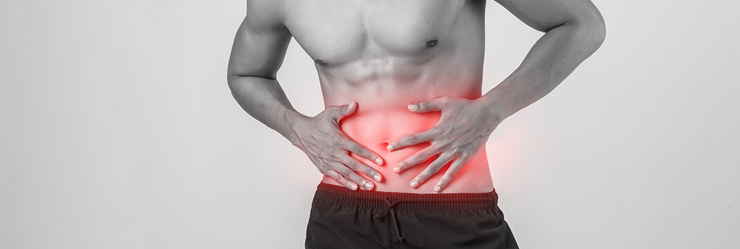
Trauma Liver and Spleen Tear
A midline incision was made from the xiphoid to below the umbilicus. Once the abdomen was opened all of the clot was evacuated. SeraSeal™ was applied to the surface of the left lobe of the liver, which had a 7 cm tear, arresting the bleeding within 3-4 seconds. All quadrants of the abdomen were packed. The packs in the lower abdominal quadrant were removed to check for associated injuries. No fecal soilage was present. The removal of the pack in the left upper quadrant was soiled with blood indicating injury to the spleen. A 2.5 cm tear to the colic surface of the spleen was located, where SeraSeal™ was applied to arrest the oozing of blood in 2-3 seconds. The right upper quadrant pack was removed with no blood saturation. Bleeding was limited to the 3 cm deep laceration to the left lobe. SeraSeal™ was applied to the base of the wound up to the surface of the lobe, achieving hemostasis in 2-3 seconds. 4-0 silk suture was used to ligate the bile ducts.
Trauma Exploring Laparotomy of the Liver
A midline incision was made from the xiphoid to below the umbilicus. Once the abdomen was opened all of the clot was evacuated. SeraSeal™ was applied to the surface of the right lobe liver fracture, arresting all bleeding within 2-5 seconds, then all quadrants of the abdomen were packed. The packs in the lower abdominal quadrant were removed to check for associated injuries. No fecal soilage was present. The removal of the packs in the left upper quadrant revealed no associated injury to the spleen. After the packs in the right upper quadrant were removed the dome of the liver rostrally was gently retracted. A gust of blood emanated from the central area indicating hepatic vein injury. Packs wee replaced. To confirm hepatic vein injury a vascular clamp was applied across the porta hepatic, which did not control the hemorrhage. Read More
Trauma Rupture Spleen
A left oblique, subcostal incision was made, beginning to the right of the midline, and proceeding obliquely outward and downward about 4 cm below the costal margin. Through dissection and mobilization, the spleen was isolated for visualization. A 4 cm longitudinal Grade 2, rupture laceration was located, and treated with SeraSeal™ deep into the splenic parenchyma to arrest the hemorrhage in 1-2 seconds. After hemostasis had occurred the wound was irrigated with sterile water and closed with absorbable horizontal mattress sutures that traversed the capsule and incorporated the parenchyma.